An image-guided biopsy of the breast is performed by taking samples of
an abnormality under some form of guidance such as ultrasound, MRI, or
mammography. A breast biopsy is performed to remove some tissue from a
suspicious area in the breast. The tissue will then be examined in the
laboratory under a microscope to determine a diagnosis. This can be performed
surgically or, more commonly, by a radiologist using a less invasive procedure
that involves a hollow needle and image-guidance. The biopsy is not designed
to remove the entire lesion, only to obtain a small sample of the abnormality
for further analysis.
How to Prepare for Interventional Breast Procedures
A consultation is generally scheduled before breast biopsies. At this time,
you will be given instructions on how to prepare and an explanation of
what to expect by a radiologist. The results of your biopsy will be sent
to your physician.
Please arrive 30 minutes before appointment time to register and complete
the necessary paperwork.
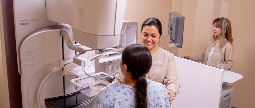
What to Expect During an Image-Guided Needle Biopsy of the Breast
A specially trained radiologist most often performs Image-guided, minimally
invasive breast procedures on an outpatient basis. Images will be obtained
from the area of interest after a local anesthetic has been injected into
the skin and more deeply into the breast to numb it. A very small nick
is made in the skin at the site where the biopsy needle is to be inserted,
and tissue samples are then removed. Typically, 3 to 12 samples are obtained,
depending on the device used. A small marker may be placed at the biopsy
site so it can be located in the future if necessary. Once the biopsy
is complete, pressure will be applied to stop any bleeding, and the opening
in the skin is covered with a dressing. No sutures are needed.
Mammogram images are obtained after the exam is complete. This procedure
is usually completed within an hour, and results will be sent to the referring
physician within 24-48 hours.